Following a week-long break, Mr. Pedersen’s junior biotechnology students wasted no time picking right up where they left off on an intricate lab: purification of GFP protein using hydrophobic interaction chromatography. Using stab cultures created during a previous lab (in which we transformed E. coli HB101 with pGLO plasmids), plates were streaked and cultures were created from the colonies produced. With the use of a lysozyme to digest the cell walls of our bacteria, a lysate was created to be purified using HIC columns.
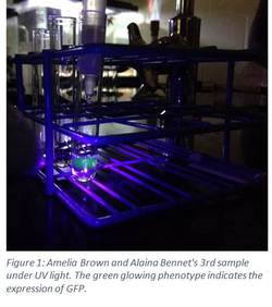
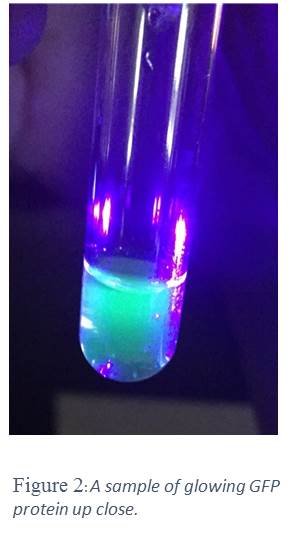
After centrifuging the lysate (which should have produced a pellet), the supernatant was loaded into the column and flowed through into a collection tube (collection tube #1). Then, wash buffer was added to the column, and the eluate was collected once again (collection tube #2). Lastly, TE buffer was added to the column, and the eluate was collected in the last tube (collection tube #3). While the first two collections did not show phenotypical evidence of GFP, the final collection did. This is due to the fact that hydrophobic proteins, such as GFP, are attracted to the resin of the HIC column. However, as the salt concentration of said resin is decreased (through wash buffer and TE buffer), the more hydrophobic proteins, in this case GFP, are able to flow through, once all other proteins have been discarded in the first two collections.
As a whole class, we considered this lab a success. Finding that we could effectively purify a protein taken from transformed bacteria weeks earlier was a huge excitement. While this lab was long and somewhat elaborate, the juniors were able to both comprehend the theory behind the lab and accurately purify GFP with little trouble. The successful samples were stored for later use/experimentation.
